Figure 1. [The normal human retina fundus]. - Webvision - NCBI
Por um escritor misterioso
Descrição
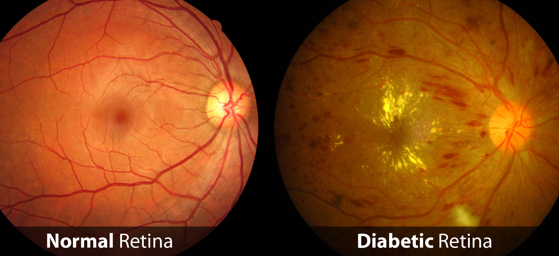
The normal human retina fundus photo shows the optic nerve (right), blood vessels and the position of the fovea (center).
![Figure 1. [The normal human retina fundus]. - Webvision - NCBI](https://journals.sagepub.com/cms/10.1177/15353702211022674/asset/images/large/10.1177_15353702211022674-fig1.jpeg)
Interpretation of anatomic correlates of outer retinal bands in optical coherence tomography - Xincheng Yao, Taeyoon Son, Tae-Hoon Kim, David Le, 2021
![Figure 1. [The normal human retina fundus]. - Webvision - NCBI](https://www.ncbi.nlm.nih.gov/books/NBK11533/bin/muller.gif)
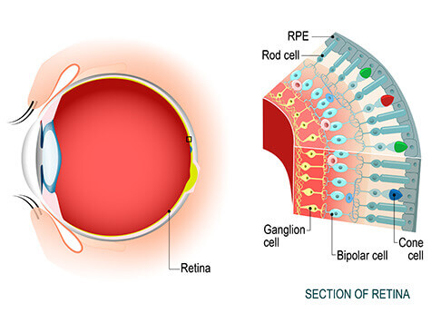
Simple Anatomy of the Retina - Webvision - NCBI Bookshelf
![Figure 1. [The normal human retina fundus]. - Webvision - NCBI](https://www.ncbi.nlm.nih.gov/books/NBK11553/bin/clinicalergf31b.jpg)
Figure 31b, [Optical coherence tomography (OCT) images]. - Webvision - NCBI Bookshelf
a) Normal fundus image. b) Pathology fundus image. c) Segmentation of
![Figure 1. [The normal human retina fundus]. - Webvision - NCBI](https://media.springernature.com/full/springer-static/image/art%3A10.1038%2Fs41467-019-12917-9/MediaObjects/41467_2019_12917_Fig1_HTML.png)
Single-nuclei RNA-seq on human retinal tissue provides improved transcriptome profiling
![Figure 1. [The normal human retina fundus]. - Webvision - NCBI](https://eophtha.com/images/uploads/15974746229665436905f37873e61580.jpg)
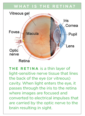
Anatomy of Retina
![Figure 1. [The normal human retina fundus]. - Webvision - NCBI](https://webvision.med.utah.edu/wp-content/uploads/2018/07/Fig-14-macula-lutea.jpg)
Simple Anatomy of the Retina by Helga Kolb – Webvision
![Figure 1. [The normal human retina fundus]. - Webvision - NCBI](https://journals.sagepub.com/cms/10.1177/15353702211022674/asset/images/large/10.1177_15353702211022674-fig8.jpeg)
Interpretation of anatomic correlates of outer retinal bands in optical coherence tomography - Xincheng Yao, Taeyoon Son, Tae-Hoon Kim, David Le, 2021
![Figure 1. [The normal human retina fundus]. - Webvision - NCBI](https://www.biorxiv.org/content/biorxiv/early/2022/02/24/2022.02.22.481546/F2.large.jpg)
Myopia alters the structural organization of the retinal astrocyte template, associated vasculature and ganglion layer thickness
![Figure 1. [The normal human retina fundus]. - Webvision - NCBI](https://www.mdpi.com/cells/cells-12-01987/article_deploy/html/images/cells-12-01987-g001.png)
Cells, Free Full-Text
de
por adulto (o preço varia de acordo com o tamanho do grupo)