Advanced Quantitative Fluorescence Microscopy to Probe the Molecular Dynamics of Viral Entry, Science Lab
Por um escritor misterioso
Descrição
Viral entry into the host cell requires the coordination of many cellular and viral proteins in a precise order. Modern microscopy techniques are now allowing researchers to investigate these interactions with higher spatiotemporal resolution than ever before. Here we present two examples from the field of HIV research that make use of an innovative quantitative imaging approach as well as cutting edge fluorescence lifetime-based confocal microscopy methods to gain novel insights into how HIV fuses to cell membranes and enters the cell.

Understanding Virus Structure and Dynamics through Molecular Simulations
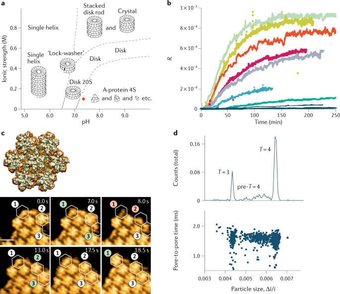
Physics of viral dynamics
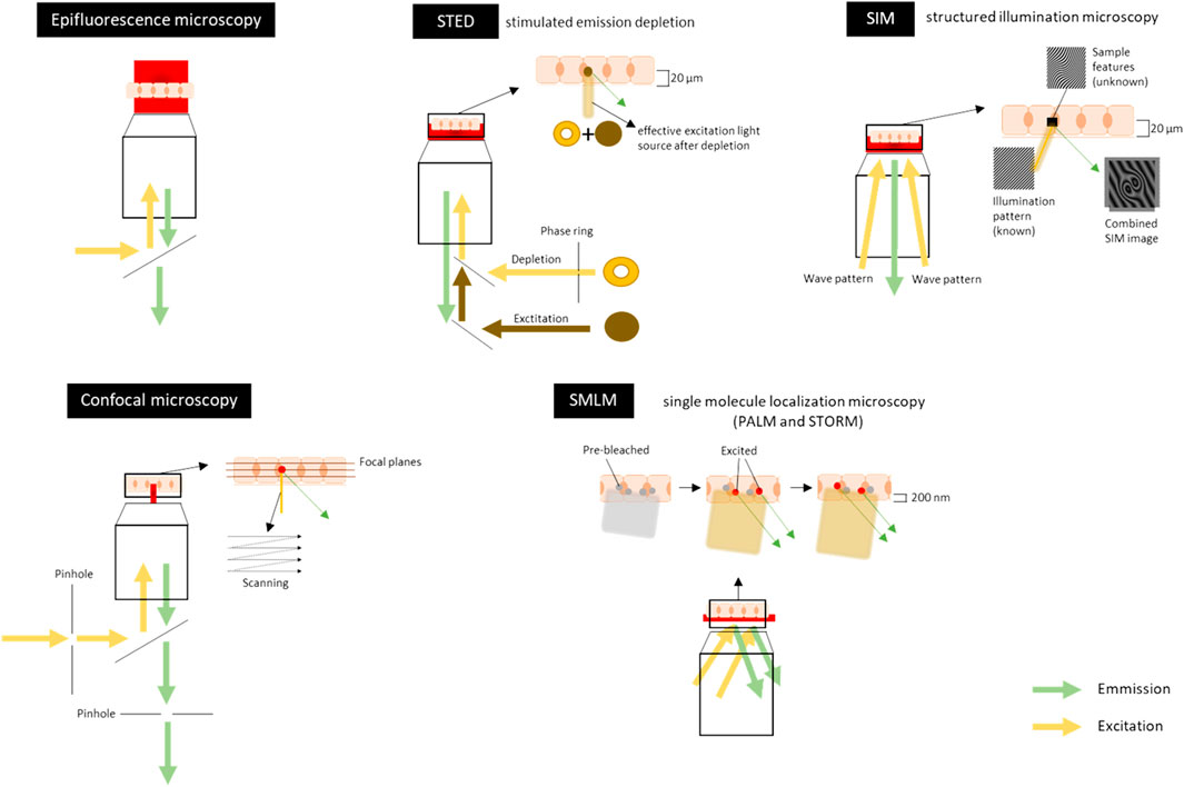
Frontiers Microscopic Visualization of Cell-Cell Adhesion Complexes at Micro and Nanoscale

Fluorescence decay curves of the purified EGFP (a) and its mutants
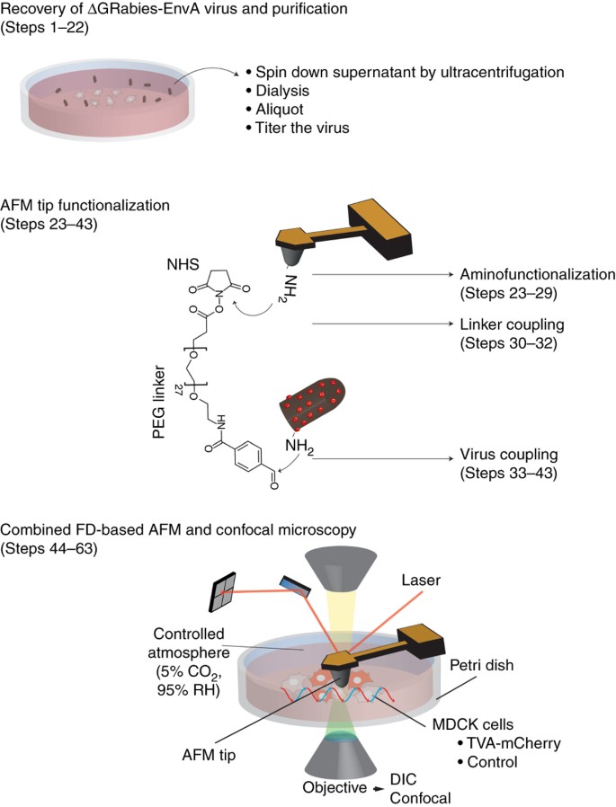
Combining confocal and atomic force microscopy to quantify single-virus binding to mammalian cell surfaces

Labeling of virus components for advanced, quantitative imaging analyses - Sakin - 2016 - FEBS Letters - Wiley Online Library
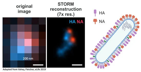
Viruses, Free Full-Text
REVIEWS ON ADVANCED MATERIALS SCIENCE
Image processing for single-virus tracking. (A) Schematic diagram of
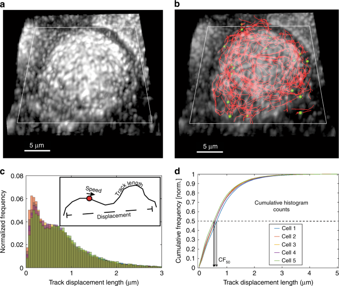
Quantitative live cell imaging reveals influenza virus manipulation of Rab11A transport through reduced dynein association

SARS-CoV-2 infection of airway cells causes intense viral and cell shedding, two spreading mechanisms affected by IL-13

Virus Entry - an overview
de
por adulto (o preço varia de acordo com o tamanho do grupo)


